
Cone Beam Imaging in Orem, UT
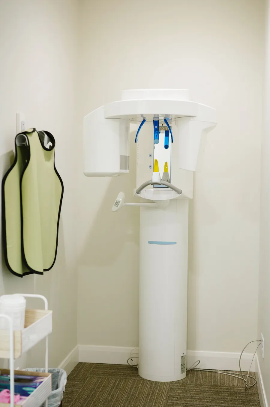
Cone Beam Imaging Explained
Cone beam computed tomography (CBCT) is a type of 3D dental X-ray. The scanner rotates around your head and captures hundreds of low-dose images, which software reconstructs into a detailed 3D model of your teeth, bone, nerves, sinuses, and jaw joints.
CBCT differs from traditional 2D X-rays by capturing depth and fine detail that flat images cannot show. It also uses a lower radiation dose than hospital-based CT scans, following the as low as reasonably achievable approach to exposure.
Benefits of Cone Beam Imaging
Comprehensive 3D detail for precise diagnosis and treatment planning.
Improved implant placement accuracy by showing bone quality and nerve location.
Better endodontic assessment by revealing hidden canals, fractures, or infections.
Clear mapping of impacted teeth and their relation to roots and sinuses.
Evaluation of jaw joints and airway for orthodontic and sleep dentistry decisions.
Fewer surprises during procedures because anatomy is visible before treatment starts.

How Cone Beam Imaging Works
- Preparation: Remove glasses, earrings, and removable metal items that could affect the image.
- Positioning: Stand or sit while the machine is adjusted so your head is stable and centered.
- Scanning: The unit rotates around your head for about 10 to 20 seconds while you stay still.
- Image Reconstruction: The system compiles a 3D image set viewable from any angle.
- Review: Our dentist reviews the results with you and explains how findings relate to your care.
Your Cone Beam Imaging Experience
Most patients find the scan easy and fast. You will not feel the X-rays and no injections or bitewing films are required. Afterward, images are available right away for immediate discussion of findings — such as bone height for a dental implant, the path of a nerve, or the presence of a missed canal.
For questions about cone beam imaging in Orem, UT, contact CrossPointe Dental at 801-224-7337.
Frequently Asked Questions About Cone Beam Imaging
No. While both create 3D images, CBCT uses a cone-shaped beam designed for dentistry and usually involves a lower radiation dose than hospital-based CT scans.
CBCT uses a low radiation dose for the amount of information it provides. Scans are prescribed only when necessary, following the ALARA principle to minimize exposure.
CBCT is often recommended for dental implants, root canal evaluations, impacted teeth, orthodontic planning, TMJ review, jaw pathology, sinus issues, and airway assessments.
The scan itself typically lasts 10 to 20 seconds. The entire appointment is usually completed within a few minutes, including positioning and image review.
Preparation is simple. Remove glasses, earrings, and metal hair accessories, and notify the team if you are or might be pregnant.
Yes. Our dentist can show you the 3D views on screen and explain how the anatomy affects your treatment choices, such as implant placement or endodontic care.
CBCT reveals bone height, width, density, and the location of nerves and sinuses. This supports accurate implant planning and helps reduce the risk of complications.